Прилад для дослідження транспорту кисню в каппілярах
Бічна панель сторінки статті
Основний зміст сторінки статті
Анотація
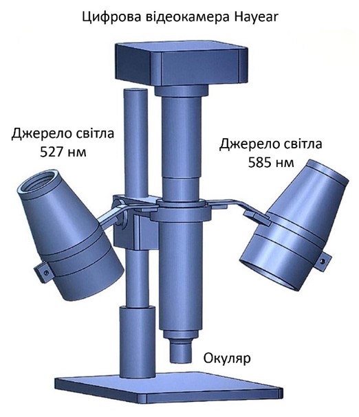
В роботі досліджується можливість візуалізації процесу передачі кисню тканинам організму через механізм капілярів. Запропоновано застосувати принцип оксиметрії на мікроскопічному рівні. Сконструйовано та виготовлено експериментальний пристрій на базі вимірювального мікроскопа та відеокамери з високою просторовою роздільною здатністю. Запропоновано й сконструйовано схеми освітлення. Описана структура приладу і його складових частин, описана ідея обробки зображень, отриманих для різних ділянок спектру. В дослідженнях використовувалось вузькосмугове освітлення в області спектральних ліній 527 нм, 585 нм та 650 нм, що формувалось за допомогою інтерференційних фільтрів з шириною смуги пропускання 10-12 нм.
Блок інформації про статтю

Ця робота ліцензується відповідно до Creative Commons Attribution 4.0 International License.
Автори, які публікуються у цьому журналі, погоджуються з наступними умовами:- Автори залишають за собою право на авторство своєї роботи та передають журналу право першої публікації цієї роботи на умовах ліцензії Creative Commons Attribution License, котра дозволяє іншим особам вільно розповсюджувати опубліковану роботу з обов'язковим посиланням на авторів оригінальної роботи та першу публікацію роботи у цьому журналі.
- Автори мають право укладати самостійні додаткові угоди щодо неексклюзивного розповсюдження роботи у тому вигляді, в якому вона була опублікована цим журналом (наприклад, розміщувати роботу в електронному сховищі установи або публікувати у складі монографії), за умови збереження посилання на першу публікацію роботи у цьому журналі.
- Політика журналу дозволяє і заохочує розміщення авторами в мережі Інтернет (наприклад, у сховищах установ або на особистих веб-сайтах) рукопису роботи, як до подання цього рукопису до редакції, так і під час його редакційного опрацювання, оскільки це сприяє виникненню продуктивної наукової дискусії та позитивно позначається на оперативності та динаміці цитування опублікованої роботи (див. The Effect of Open Access).
Посилання
K. von Vierordt, “Die Anwendung des Spectralapparates zur Photometrie der Absorptionsspektren und zur quantitativen chemischen Analyse.” Tübingen: Laupp, 1872, URL: https://vlp.mpiwg-berlin.mpg.de/people/data?id=per166.
B. L. Horecker, “The absorption spectra of hemoglobin and its derivatives in the visible and near infra-red regions,” J Biol Chem, vol. 148, no. 1, pp. 173–183, 1943, DOI: https://doi.org/10.1016/S0021-9258(18)72329-6.
G. A. Millikan, “The Oximeter, an Instrument for Measuring Continuously the Oxygen Saturation of Arterial Blood in Man,” Rev. Sci. Instrum., vol. 13, no. 10, pp. 434–444, Oct. 1942, DOI: https://doi.org/10.1063/1.1769941.
M. W. Wukitsch, M. T. Petterson, D. R. Tobler, and J. A. Pologe, “Pulse oximetry: Analysis of theory, technology, and practice,” J. Clin. Monit., vol. 4, no. 4, pp. 290–301, Oct. 1988, DOI: https://doi.org/10.1007/BF01617328.
H.-W. M. Breuer, H. Groeben, J. Breuer, and H. Worth, “Oxygen saturation calculation procedures: A critical analysis of six equations for the determination of oxygen saturation,” Intensive Care Med., vol. 15, no. 6, Sep. 1989, DOI: https://doi.org/10.1007/BF00261498.
J. W. Severinghaus and Y. Honda, “History of blood gas analysis. VII. Pulse oximetry,” J. Clin. Monit., vol. 3, no. 2, pp. 135–138, Apr. 1987, DOI: https://doi.org/10.1007/BF00858362.
M. L. J. Landsman, N. Knop, G. Kwant, G. A. Mook, and W. G. Zijlstra, “A fiberoptic reflection oximeter,” Pflügers Arch. Eur. J. Physiol., vol. 373, no. 3, pp. 273–282, Mar. 1978, DOI: https://doi.org/10.1007/BF00580835.
C. G. Ellis, M. L. Ellsworth, R. N. Pittman, and W. L. Burgess, “Application of image analysis for evaluation of red blood cell dynamics in capillaries,” Microvasc. Res., vol. 44, no. 2, pp. 214–225, Sep. 1992, DOI: https://doi.org/10.1016/0026-2862(92)90081-Y.
C.-C. Wu et al., “Accuracy evaluation of RBC velocity measurement in nail-fold capillaries,” Microvasc. Res., vol. 81, no. 3, pp. 252–260, May 2011, DOI: https://doi.org/10.1016/j.mvr.2011.01.003.
A. F. Fercher and J. D. Briers, “Flow visualization by means of single-exposure speckle photography,” Opt. Commun., vol. 37, no. 5, pp. 326–330, Jun. 1981, DOI: https://doi.org/10.1016/0030-4018(81)90428-4.
H. Fujii, K. Nohira, Y. Yamamoto, H. Ikawa, and T. Ohura, “Evaluation of blood flow by laser speckle image sensing Part 1,” Appl. Opt., vol. 26, no. 24, p. 5321, Dec. 1987, DOI: https://doi.org/10.1364/AO.26.005321.
J. D. Briers, “Laser speckle contrast analysis (LASCA): a nonscanning, full-field technique for monitoring capillary blood flow,” J. Biomed. Opt., vol. 1, no. 2, p. 174, 1996, DOI: https://doi.org/10.1117/12.231359.
F. C. Delori, “Noninvasive technique for oximetry of blood in retinal vessels,” Appl. Opt., vol. 27, no. 6, p. 1113, Mar. 1988, DOI: https://doi.org/10.1364/AO.27.001113.
S. H. Hardarson, “Retinal Oximetry,” Univ Iceland, 2012.
C. Criveanu et al., “Capillaroscopy – a valuable tool for the assessment of vascular impairment in systemic sclerosis,” Rom. J Rheumatol, vol. 25, no. 3, pp. 112–116, 2016. URL: https://rjr.com.ro/articles/2016.3/RJR_2016_3_Art-02.pdf
M. S. Molebna, “Doslidzhennia spektralnoi mikroskopichnoi oksymetrii krovi”. Zvit pro naukovo-doslidnytsku robotu MAN. Viddilennia tekhnichnykh nauk. [‘Studies of spectral microscopic blood oxymetry’. Report on R&D at Small Academy of Sciences],” Kyiv (manuscript, in Ukrainian), 2021.
V. Molebny and M. Molebna, “Capillary oxymeter,” in European Conferences on Biomedical Optics (ECBO 2021), 2021.
V. Molebny and M. Molebna, “Instrument for visualization of oxygen transport in nailfold capillaries as an aid in diagnosing oxygen-deficiency diseases,” in Euro Asia 8th International Congress on Applied Sciences, 2021.
V. Molebny and M. Molebna, “Illumination for Capillary Oxymeter,” in OSA Imaging and Applied Optics Congress, 2021.
“LSR-3005 — Optotune.” URL: https://www.optotune.com/lsr-3005.
“SC-5-HF High frequency resonant optical scanner.” URL: http://www.eopc.com/sc5.html.
V. Molebny and M. Molebna, “Spectral narrow-band illumination of the object field in capillary oximetry,” in The Fifth International Conference on Biological Information and Biomedical Engineering (BIBE 2021), 2021.